An electroencephalogram (EEG) is a test that measures and records the electrical activity of your brain. Special sensors (electrodes) are attached to your head and hooked by wires to a computer. The computer records your brain's electrical activity on the screen or on paper as wavy lines. Certain conditions, such as seizures, can be seen by the changes in the normal pattern of the brain's electrical activity.
Why It Is Done?
An electroencephalogram (EEG) may be done to:
- Diagnose epilepsy and see what type of seizures are occurring. EEG is the most useful and important test in confirming a diagnosis of epilepsy.
- Check for problems with loss of consciousness or dementia.
- Help find out a person's chance of recovery after a change in consciousness.
- Find out if a person who is in a coma is brain-dead.
- Study sleep disorders, such as narcolepsy.
- Watch brain activity while a person is receiving general anesthesia during brain surgery.
- Help find out if a person has a physical problem (problems in the brain, spinal cord, or nervous system) or a mental health problem.
How To Prepare?
Before the day of the electroencephalogram (EEG) test, tell your doctor if you are taking any medicines. Your doctor may ask you to stop taking certain medicines (such as sedatives and tranquilizers, muscle relaxants, sleeping aids, or medicines used to treat seizures) before the test. These medicines can affect your brain's usual electrical activity and cause abnormal test results.
Do not eat or drink foods that have caffeine (such as coffee, tea, cola, and chocolate) for 8 hours before the test.
Since the electrodes are attached to your scalp, it is important that your hair be clean and free of sprays, oils, creams, and lotions. Shampoo your hair and rinse with clear water the evening before or the morning of the test. Do not put any hair conditioner or oil on after shampooing.
To find certain types of abnormal electrical activity in the brain, you may have to be asleep during the recording. You may be asked not to sleep at all the night before the test or to sleep less (about 4 or 5 hours) by going to bed later and getting up earlier than usual. If your child is going to be tested, try to keep him or her from taking naps just before the test. If you know that you are going to have a sleep-deprived EEG, plan to have someone drive you to and from the test.
How It Is Done?
An electroencephalogram (EEG) may be done in a hospital or in a doctor's office by an EEG technologist. The EEG record is read by a doctor who is specially trained to diagnose and treat disorders affecting the nervous system (neurologist).
Pulmonary function tests are a group of tests that measure how well the lungs take in and release air and how well they move gases such as oxygen from the atmosphere into the body's circulation.
How the Test is Performed?
In a spirometry test, you breathe into a mouthpiece that is connected to an instrument called a spirometer. The spirometer records the amount and the rate of air that you breathe in and out over a period of time.
For some of the test measurements, you can breathe normally and quietly. Other tests require forced inhalation or exhalation after a deep breath.
How to Prepare for the Test?
Do not eat a heavy meal before the test. Do not smoke for 4 - 6 hours before the test. You'll get specific instructions if you need to stop using bronchodilators or inhaler medications. You may have to breathe in medication before the test.
How the Test Will Feel?
Since the test involves some forced breathing and rapid breathing, you may have some temporary shortness of breath or light-headedness. You breathe through a tight-fitting mouthpiece, and you'll have nose clips.
Why the Test is Performed?
Pulmonary function tests are done to:
- Diagnose certain types of lung disease (especially asthma, bronchitis, and emphysema)
- Find the cause of shortness of breath
- Measure whether exposure to contaminants at work affects lung function
It also can be done to:
- Assess the effect of medication
- Measure progress in disease treatment
Spirometry measures airflow. By measuring how much air you exhale, and how quickly, spirometry can evaluate a broad range of lung diseases.
Lung volume measures the amount of air in the lungs without forcibly blowing out. Some lung diseases (such as emphysema and chronic bronchitis) can make the lungs contain too much air. Other lung diseases (such as fibrosis of the lungs and asbestosis) make the lungs scarred and smaller so that they contain too little air.
Testing the diffusion capacity (also called the DLCO) allows the doctor to estimate how well the lungs move oxygen from the air into the bloodstream.
Pulmonary function tests are a group of tests that measure how well the lungs take in and release air and how well they move gases such as oxygen from the atmosphere into the body's circulation.
Gastroscopy is also Known as
- EGD
- Esophagogastroduodenoscopy
- Esophagoscopy
- Peroral Endoscopy
- Upper Endoscopy
What is gastroscopy?
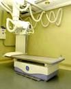 |
Radiography is the term for a general x-ray exam that captures clear, precise images using radiation.
Radiation, a form of energy, exists in nature and emanates from the atmosphere and earth. As with many naturally-occurring substances, radiation, in moderation, is considered harmless.
X-ray beams can pass through the human body. When they strike a detector, they produce a picture.
Traditional film-based exams have been replaced by digital imaging in many cases. Digital radiography requires no film processing. Test results can be viewed seconds after the exposure is made. |
How is a gastroscopy performed?
X-rays help to diagnose a wide variety of conditions including bone injuries, infections, arthritis and cancer. A doctor can get a detailed view of the spine, fingers, toes, abdomen, urinary tract, gastrointestinal system, chest, ribs, skull, sinuses, facial bones and other specific areas of the body.
Why is gastroscopy useful?
Depending on the part of the body being x-rayed, the patient may be asked to lie on a table, sit, or stand while the images are taken. The patient will be given a lead apron to wear for protection of parts of the body that are not to be x-rayed.
Some types of x-ray exams require the use of a “contrast medium” that is either injected or taken orally in order to allow the doctor to see inside blood vessels or the urinary tract.
The exam usually takes 10-45 minutes to complete.
Women who are, or may be, pregnant or are breastfeeding, must alert their doctor and the technologist if they are being scheduled for an x-ray procedure.
X-rays are among Greenwich Hospital's broad range of diagnostic and interventional radiology services.
Radiology services are generally pain-free, non-invasive and available to both outpatients and inpatients.
Can gastroscopy be used to examine other parts of the body?
X-rays help to diagnose a wide variety of conditions including bone injuries, infections, arthritis and cancer. A doctor can get a detailed view of the spine, fingers, toes, abdomen, urinary tract, gastrointestinal system, chest, ribs, skull, sinuses, facial bones and other specific areas of the body.
How far can a gastroscope see?
X-rays help to diagnose a wide variety of conditions including bone injuries, infections, arthritis and cancer. A doctor can get a detailed view of the spine, fingers, toes, abdomen, urinary tract, gastrointestinal system, chest, ribs, skull, sinuses, facial bones and other specific areas of the body.
Are there other uses for gastroscopy?
X-rays help to diagnose a wide variety of conditions including bone injuries, infections, arthritis and cancer. A doctor can get a detailed view of the spine, fingers, toes, abdomen, urinary tract, gastrointestinal system, chest, ribs, skull, sinuses, facial bones and other specific areas of the body.
Why doesn't my doctor just send me for an X-ray?
X-rays help to diagnose a wide variety of conditions including bone injuries, infections, arthritis and cancer. A doctor can get a detailed view of the spine, fingers, toes, abdomen, urinary tract, gastrointestinal system, chest, ribs, skull, sinuses, facial bones and other specific areas of the body.
Is gastroscopy safe?
X-rays help to diagnose a wide variety of conditions including bone injuries, infections, arthritis and cancer. A doctor can get a detailed view of the spine, fingers, toes, abdomen, urinary tract, gastrointestinal system, chest, ribs, skull, sinuses, facial bones and other specific areas of the body.
Definition
Audiometry is the testing of a person's ability to hear various sound frequencies. The test is performed with the use of electronic equipment called an audiometer. This testing is usually administered by a trained technician called an audiologist.
Description
A trained audiologist (a specialist in detecting hearing loss) uses an audiometer to conduct audiometry testing. This equipment emits sounds or tones, like musical notes, at various frequencies, or pitches, and at differing volumes or levels of loudness. Testing is usually done in a soundproof testing room.
The person being tested wears a set of headphones that blocks out other distracting sounds and delivers a test tone to one ear at a time. At the sound of a tone, the patient holds up a hand or finger to indicate that the sound is detected. The audiologist lowers the volume and repeats the sound until the patient can no longer detect it. This process is repeated over a wide range of tones or frequencies from very deep, low sounds, like the lowest note played on a tuba, to very high sounds, like the pinging of a triangle. Each ear is tested separately. It is not unusual for levels of sensitivity to sound to differ from one ear to the other.
A second type of audiometry testing uses a headband rather than headphones. The headband is worn with small plastic rectangles that fit behind the ears to conduct sound through the bones of the skull. The patient being tested senses the tones that are transmitted as vibrations through the bones to the inner ear. As with the headphones, the tones are repeated at various frequencies and volumes.
The results of the audiometry test may be recorded on a grid or graph called an audiogram. This graph is generally set up with low frequencies or tones at one end and high ones at the other end, much like a piano keyboard. Low notes are graphed on the left and high notes on the right. The graph also charts the volume of the tones used; from soft, quiet sounds at the top of the chart to loud sounds at the bottom. Hearing is measured in units called decibels. Most of the sounds associated with normal speech patterns are generally spoken in the range of 20-50 decibels. An adult with normal hearing can detect tones between 0-20 decibels.
Speech audiometry is another type of testing that uses a series of simple recorded words spoken at various volumes into headphones worn by the patient being tested. The patient repeats each word back to the audiologist as it is heard. An adult with normal hearing will be able to recognize and repeat 90-100% of the words.