Radiology
Doctors Diagnostic radiology Services covers a broad range of diagnostic tests from chest X-rays to gastrointestinal (GI) series to intravenous pyelograms. Some of the tests, such as chest X-rays or simple bone X-rays, require no special diet or preparation. Other tests, such as the upper GI or barium enema, require preparation through special diets one or several days before the exam. Doctors Diagnostic's Radiology Department will give you sample menus and information on preparing for the test.
Ultrasound
What is an ultrasound?
Ultrasound (also known as sonogram) is a safe, state-of-the-art exam. High frequency sound waves are used to produce real-time visual images that differentiate between the body's soft tissues and its fluid-filled structures. Doppler ultrasound can also detect motion, such as the movement of blood cells.
Unlike x-rays and CT scans, ultrasound does not use radiation and, therefore, may be used safely during pregnancy.
Why would I need an ultrasound?
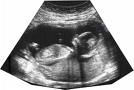 |
Ultrasound allows a physician to view and evaluate veins, arteries and blood flow in a person's neck, arms, abdomen and legs. In pregnancy, ultrasound can help determine fetal age and anatomical development. It also may be used to screen a fetus at risk for Down syndrome in the first trimester. Ultrasound technology is helpful too in the area of breast health, when a questionable mammogram finding requires more detailed exploration. |
What to Expect?
A clear water-based gel is applied to the part of the body being scanned. This reduces small amounts of air that can interfere with imaging.
A smooth hand-held device called a transducer is gently rubbed across the part of the body being examined. Sound waves generated from within the instrument enter the body and returning echoes are transferred back to a computer.
The reflected sound waves are used to produce live images on a monitor and allow real-time imaging of an area of interest.
Since the images are generated in real time, they can be used to show the structure and movements of internal organs and muscles. During this time, the patient simply lies still. No special diet or preparation is needed beforehand.
Radiology services are generally pain-free, non-invasive and available to both outpatients and inpatients.
Bone Density Testing – Detecting Osteoporosis
What is a bone density test?
A bone mineral density test measures bone strength and quality. It can detect as little as one to three percent bone loss with one-tenth the radiation exposure of a chest x-ray. (As comparison, a general x-ray is unable to detect osteoporosis until bone loss reaches 30 percent.
Why would I need a bone density test?
A bone density test is used to diagnose osteoporosis and evaluate risk for fracture. This allows for prevention and early treatment of bone loss. Periodic testing helps the physician track the success of treatment and allows for adjustments before symptoms develop.
What to Expect?
The test takes about 20 minutes. The initial test is used as a reference for comparison with future measurements.
Understanding Osteoporosist
Osteoporosis is a condition causing bones to become weak and brittle. This can lead to fractures of the spine, hip, ankle and wrist from simple falls that would not impact people with healthy bones. Collapsed vertebrae due to osteoporosis can result in severe back pain, loss of height and stooped posture or other spinal deformities.
Osteoporosis most commonly appears in women in the first five to ten years following menopause, and advances with age.
Osteoporosis doesn't usually produce symptoms until a fracture occurs, often resulting from a fall.
Women who are, or think they may be, pregnant should not have any type of bone density exam.
Bone density testing is among Greenwich Hospital's broad range of diagnostic and interventional radiology services.
Radiology services are generally pain-free, non-invasive and available to both outpatients and inpatients.
Radiography
What is radiography?
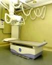 |
Radiography is the term for a general x-ray exam that captures clear, precise images using radiation.
Radiation, a form of energy, exists in nature and emanates from the atmosphere and earth. As with many naturally-occurring substances, radiation, in moderation, is considered harmless.
X-ray beams can pass through the human body. When they strike a detector, they produce a picture.
Traditional film-based exams have been replaced by digital imaging in many cases. Digital radiography requires no film processing. Test results can be viewed seconds after the exposure is made. |
Why do I need an x-ray?
X-rays help to diagnose a wide variety of conditions including bone injuries, infections, arthritis and cancer. A doctor can get a detailed view of the spine, fingers, toes, abdomen, urinary tract, gastrointestinal system, chest, ribs, skull, sinuses, facial bones and other specific areas of the body.
What to Expect?
Depending on the part of the body being x-rayed, the patient may be asked to lie on a table, sit, or stand while the images are taken. The patient will be given a lead apron to wear for protection of parts of the body that are not to be x-rayed.
Some types of x-ray exams require the use of a “contrast medium” that is either injected or taken orally in order to allow the doctor to see inside blood vessels or the urinary tract.
The exam usually takes 10-45 minutes to complete.
Women who are, or may be, pregnant or are breastfeeding, must alert their doctor and the technologist if they are being scheduled for an x-ray procedure.
X-rays are among Greenwich Hospital's broad range of diagnostic and interventional radiology services.
Radiology services are generally pain-free, non-invasive and available to both outpatients and inpatients.
CT Scan
What is an Ultrasound?
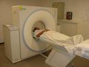 |
A computed tomography (CT) scan uses x-rays and a sophisticated computer to view specific parts of the body's anatomy in great detail. It is a very common imaging exam.
Unlike a traditional x-ray, where the radiation beam comes from a stationary or non-moving source, a CT scan is created by moving the x-ray beam around the patient to obtain horizontal and vertical cross-sectional views. Spiral or helical CT scans can capture three-dimensional images.
|
Ultrasound allows a physician to view and evaluate veins, arteries and blood flow in a person's neck, arms, abdomen and legs. In pregnancy, ultrasound can help determine fetal age and anatomical development. It also may be used to screen a fetus at risk for Down syndrome in the first trimester. Ultrasound technology is helpful too in the area of breast health, when a questionable mammogram finding requires more detailed exploration. |
Why would I need a CT scan?
A CT scan allows the physician to see various angles of a particular structure such as the brain, the heart or joints inside the body. It is sometimes used to diagnose coronary artery disease
What to Expect?
The CT scanner is a large doughnut shaped machine. The patient lies on a table with the part of the body to be examined positioned within the scanner opening. The table moves slowly and periodically during the procedure. A whirring or whooshing sound may be heard as the scan is performed.
It's important for the patient to lie still during the exam to make the images as clear as possible. The patient will be able to speak to the technologist performing the exam through a built-in intercom system at all times.
Depending on the part of the body being scanned, a contrast medium, administered orally or by injection into a vein, may be required. For other organs, fasting may be required.
A CT scan usually takes about 20-30 minutes.
Female patients who may be pregnant or are breastfeeding should discuss this with the physician prior to scheduling and with the technologist prior to the scan.
Radiology services are generally pain-free, non-invasive and available to both outpatients and inpatients.
Breast Imaging – Mammogram
What is Breast Imaging?
.jpg) |
Although patients may be familiar with routine mammograms, many women don't realize that these are only one of several breast imaging procedures, each serving a different purpose.
A screening mammogram is a detailed x-ray of the breast that can detect cancerous or precancerous areas before a lump is found on physical exam.
|
A diagnostic mammogram is offered to women with breast symptoms such as a lump, nipple discharge or skin change. They are also used for women who have previously had an abnormal screening and for women with implants.
Breast ultrasound is offered to women with dense breasts or to evaluate lumps that have been detected either on physical examination or by mammography.
Needle localization can mark abnormal imaging findings for surgeons to guide excisional biopsy.
Image-guided core biopsy is a minimally invasive needle biopsy performed under local anesthesia.
Computer Aided Diagnosis (CAD) is used to help improve early detection of breast cancer. |
Why do I need a mammogram?
A mammogram is the single best test for detecting breast cancer in its earliest stages.
Protecting Women's Good Health
A clear water-based gel is applied to the part of the body being scanned. This reduces small amounts of air that can interfere with imaging.
A smooth hand-held device called a transducer is gently rubbed across the part of the body being examined. Sound waves generated from within the instrument enter the body and returning echoes are transferred back to a computer.
The reflected sound waves are used to produce live images on a monitor and allow real-time imaging of an area of interest.
Since the images are generated in real time, they can be used to show the structure and movements of internal organs and muscles. During this time, the patient simply lies still. No special diet or preparation is needed beforehand.
Radiology services are generally pain-free, non-invasive and available to both outpatients and inpatients
|
| Related Services |
Cardiology |
|
Other Services |
|
 |
 |
|